WELCOME..... TO .....HIGH SCHOOL BIOLOGY BLOG !!!

Monday, July 9, 2012

Kidney, Osmoregulation and Urine Formation

KIDNEY


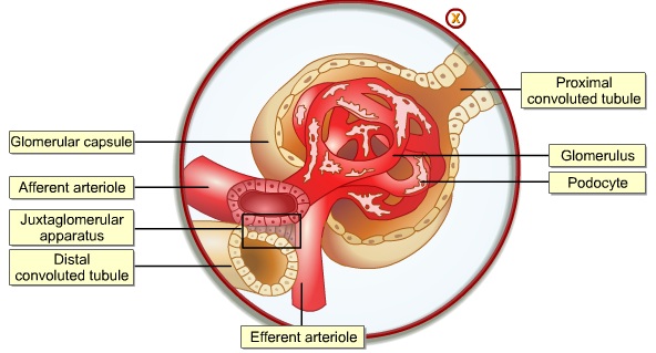
 1.      Each kidneys contains approximately one to two million tiny functional units (nephrons).


2.      Each nephron consists of:
      -          Bowman’s capsule
      -          proximal convoluted tubule
      -          loop of Henle
      -          distal convoluted tubule
      -          collecting duct.


3.      Each nephron tubule - is extensively surrounded by a capillary network.
4.      Blood:
      -          enters kidney through renal artery
      -          leaves through renal vein.
      -          afferent arteriole carries blood à to glomerulus
      -          efferent arteriole carries blood ßout of  glomerulus.


5.      High blood pressure (in glomerulus) forces blood plasma à into Bowson’s capsule due to:
  à renal artery is short + close to aorta.
  à diameter of the efferent arteriole is smaller than afferent arteriole.

URINE FORMATION

1.      Three processes involved in the urine formation are:
      -          ultrafilteration,
      -          reabsorption
      -          secretion.

2.      Ultrafilteration:
      -          a process
      -          blood plasma is forced out from the glomerulus
      -          into the Bowman’s capsule
      -          due to high hydrostatic pressure.


3.      Glomerular filtrate contains:
      -          ions,
      -          water
      -          nutrients
      -          nitrogenous waste.

4.      Reabsorption of  glucose +  amino acids
      -          occurs at the proximal convoluted tubule
      -          an active transport.


5.      Reabsorption of water:
      -          occurs at descending limb of loop of Henle.


6.      Secretion of hydrogen ions (H+), creatinine, and toxic waste
      -          occurs at distal convoluted tubule.


7.      Urea is excreted, while water, some urea and sodium ions (Na+)
      -          reabsorbed at collecting duct.
      -          composition of urine =  ions, urea, water, toxins + creatinine.
     

OSMOREGULATION

1.      Osmoreceptors (in hypothalamus) - constantly monitor the amount of water in blood.
2.      ADH:
  è released by the posterior lobe of pituitary
  è regulates the reabsorption of the water
  è by kidney.


3.      When osmotic pressure (OP↑) in blood increase:
  è ADH is secreted
  è increases permeability of collecting duct + distal tubule
  è to reabsorb more water.
  è Consequently, less more concentrated urine is produces.

4.      When osmotic pressure (OP↓) decreases:
  è adrenal cortex releases aldosterone (2nd  hormone)
  è stimulates distal convoluted tubule +  collecting duct
  è to reabsorb more sodium ions (Na+)
  è thru active transport,
  è increases blood volume ↑.
  è regulates blood concentration + volume.

5.      rennin-angiotensin mechanismà trigger the release of aldosterone.

6.      Negative feedback mechanism:
  è a system
  è which brings an imbalance back to normal
  è by responding to a condition it controls.

7.      Blood pH is maintained - between 7.35 to 7.45.
      -     relative exchange of bicarbonate (HCO3-) + hydrogen ions (H+)
      -     between blood +  tubules
      -     help adjust acid-base balance of blood.


8.      When blood pH ↓ decreases:
      -          H+ are secreted into distal tubule
      -          while HCO3- are reabsorbed,
      -          resulting in blood pH returning to normal.

9.      When blood pH ↑ increases:
      -          HCO3- will be removed
      -          from blood àinto distal tubule.
      -          few H+ à move into blood capillary.

10.  Urine pH vary between 4.5 -  8.2 (normal pH = 6.0).


LIVER II

1.      Cori cycle à involves liver + skeletal muscle.
2.      During strenuous exercise:
      è glucose (in muscle cells)
      è converted to lactate (produces 2ATP)
      è transported to liver (by blood stream) to be metabolized.

3.      In liver:
      è lactate is oxidized to pyruvate
      è then to glucose (using ATP).
      è transported back to the muscle (via bloodstream).


4.      Ornithine cycle (in mitochondria):
      è produces amino group (NH3)
      è from deamination process
      è NH3 + CO2 à carbamoyl phosphate.
      è carbamoyl phosphate + ornithine à citruline (amino acid).
      è 2nd amino group (aspartate) à enters the ornithine cycle.
      è citruline is converted back by a multistep reaction to ornithine
          (with the formation of urea).
      è ornithine + carbarmoyl phosphate à repeat the cycle


Friday, July 6, 2012

LIVER I


1.      Liver - made up of thousands of lobules.


2.      Each lobule - consists of rows of liver cells (hepatocytes).
3.      Sinusoids:
      - channels where blood flows
      - from the hepatic artery
      - into the hepatic vein.
4.      Hepatic vein:
      - carries deoxygenated blood + nutrients
      - to the heart.
5.      Hepatic portal vein:
      - carries blood-containing nutrients
      - from intestine
      - into lobules.
6.      Canalicluli:
      - where bile secreted by the liver cells flows
      - into bile duct.


7.      Liver performs many important functions, all of which contribute to homeostasis:
      i.        Carbohydrate + protein metabolism
      ii.      site of synthesis of lipid (from excess glucose).
      iii.    detoxification - where drugs, toxins and poisons
             (taken in the diet/produced by bacteria)
             biochemically broken down + rendered harmless.
      iv.    production of bile + plasma proteins,
      v.      storage site of ions, minerals + vitamins
      vi.    generation of heat

8.      In the liver: glucose is oxidized à to CO2 + H2O.
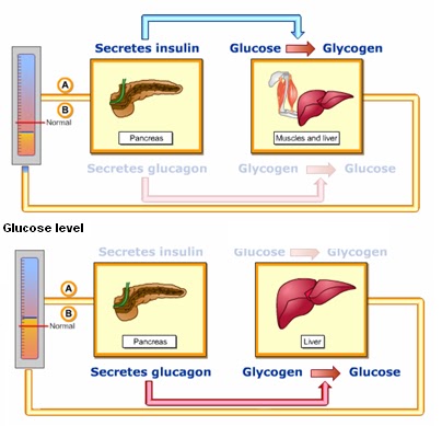
9.      In the liver cells (stimulated by insulin)
      à excess glucose is converted to glycogen (glycogenesis),
10.  When the levels of blood glucose drop
      à glycogen is converted to glucose (presence of glycagon).
11.  When glycogen ↓ in the liver cells is depleted and glucose↓ level fall
      à glucose is made from other non-carbohydrate sources = gluconeogenesis.

Thursday, March 8, 2012

U6ScBeta 2012 - rat dissection


One for me...one for you ...and one for.....

Get set!...ready!... cuuuuuuuuut....!!!

Give me...give me...please...please...please.......!

I told you....I'm going to skin you alive.....!

This is how you skin a RAT!!!!

I think the belly button... is around here.... somewhere......!!!

Hah!.....I've found it...the stolen chicken fat...!!!

Hey....where is the...?

Aiyah!!!.....this rat has babies in it...!!!!


Display of rat digestive organs.
Ouch!!!..you've broke my heart...
Bloody bleeding rat...!!!
Hey!....I've found something here...!
Sorry!...no peeping...it's a secret mission....!
Sob...sob...I thought I have to cut only....


Tuesday, March 6, 2012

U6scZeta 2012 - Rat Dissection

2012
Here kitty kitty... here kitty kitty... nice kitty...!

Here....take it ...make sure it's properly cooked!

Here you are....this is a big one... specially kept.. for U!

Like this....massage here...and a little bit there...that's the way!

Help!!!.. I'm stuck!!!...

Yes...snip snip here....and... snip snip there....!

Oh no... not there! It's tickling there.....haha..hoho..haha....

Help!...I've been violated!!!!

Bevy of maidens with their presentations on... rat dissection!

Sorry!.. you have been exposed........

Now... I know... the rubbish inside you.....!

Sunday, November 6, 2011

Homeostasis ll

1.   The pancreas
- functions as exocrine + endocrine gland.
- has 2 groups of cell in the pancreas.
- α & β-cells in the islet of Langerhans play (major role in blood glucose concdntration).
- α-cells - secrete glucagons
- β-cells - secrete insulin into blood capillaries.

α & β-cells in the islet of Langerhans


2.   Blood glucose level rises↑à due to carbohydrate breakdown.
3.   Insulin + glucagons have opposite effects.
4.   Insulin:
-  secreted by β-cells (when glucose↑ level increases).
-  target organs = liver + muscle tissues.
-  convert glucose à to glycogen.
5.   Glucagon:
-  secreted by α-cells (when the glucose↓ level decreases)
-  Target organ = liver cells.
-  convert glycogen à glucose.

Pancreas response to different level of glucose


6.   Ingestion of glucose à increase insulin level ↑.
7.   2 types of diabetes: Type I & Type II.
Blood glucose & insulin level in normal & diabetic person.


8.   Formation of tissues fluid is a physical process.
9.   At arteriol end:  
-  smaller blood plasma content is forced out
-  when hydrostatic pressure (HP) > osmotic pressure (OP).
10. At venous end:
-  tissue fluid re-enters capillary
-  when osmotic pressure (OP) > hydrostatic pressure (HP).